
Bimaxillary Immediate Loading: When Planning Meets Clinical Reality
It has been a while since I last wrote something for the blog. The truth is that life at the clinic has been moving at an incredible pace. These past two months have been intense in every possible way — a lot of work, many challenges and, above all, an enormous learning process.
But I finally feel that we are settling into a good rhythm. There is some light at the end of the tunnel.
Yesterday, we treated one of those cases that reminds us exactly why Implant Dentistry remains so challenging: a full maxillomandibular rehabilitation with bimaxillary immediate loading.
A socially unsustainable situation
The patient came to us following the recommendation of another patient who had previously undergone treatment at our clinic.
Even during a normal conversation, the mobility of her teeth was striking. They were so
mobile that they almost appeared to be coming out of her mouth as she spoke.
It was a functionally and socially unsustainable situation.
The first thing we did was what we should always do, even when the clinical picture seems obvious: evaluate every single tooth individually.
We assessed the periodontal condition, attachment loss, mobility, tooth position, and the realistic possibility of preserving each tooth through periodontal therapy.
Even on our most optimistic days when it comes to saving teeth, it quickly became clear that this would not be possible in this case.
The attachment loss was extraordinary.
In the first and second quadrants, there was actually an interesting situation: when the patient smiled, the gingival margins appeared to be in a relatively acceptable position. But that appearance was misleading. The migration of the junctional epithelium concealed an enormous loss of periodontal attachment.
The conclusion in the mandible was essentially the same.
There was no predictable way of maintaining this dentition.
So the question changed from:
“Which teeth can we save?”
to:
“How are we going to rehabilitate this patient?”
Teeth first. Implants second.
Before thinking about implants, we performed a functional and aesthetic analysis.
We imported the case into SmileCloud and began by determining two fundamental positions: where the maxillary central incisor should end and where the first molars should be positioned.
For me, this is one of the most important parts of a maxillomandibular rehabilitation.
The central incisor drives the entire rehabilitation.
fig
However, because virtually all the teeth in this patient were out of position, it was equally important to determine where teeth 16 and 26 should be located.
fig
We like to use concepts derived from the orthodontic literature as references. In a Class I normal occlusion, one of the parameters we look for is an intermolar distance of approximately 31–35 mm, measured between the palatal cusps of the maxillary first molars.
In a patient whose teeth were so severely displaced, this reference became particularly useful in helping us establish the appropriate positions of 16 and 26.
Together with the position of the central incisor, this allowed us to begin reconstructing the architecture of the maxillary arch.
Once the maxillary position had been established, we knew that the vertical dimension would then have to be organised through its relationship with the mandibular arch.
Only then did we really start thinking about implants.
From SmileCloud to Exocad and coDiagnostiX ( sofia)
Once we had established in SmileCloud where we wanted the teeth to end up, we needed to transfer that information into our planning workflow.
From SmileCloud, we can export an STL containing all the teeth in the positions we have digitally determined.
This is an important step.
We are no longer dealing simply with a visual representation of the smile we would like to achieve. We now have a three-dimensional object that can be transferred between different software platforms.
The STL of this digital tooth setup is then imported into Exocad.
In Exocad, we bring two different worlds together.
On one side, we have the patient's original STL, representing the initial clinical situation.
On the other, we have the STL containing the teeth in the positions established in SmileCloud.
By bringing both datasets into the same coordinate system, we can reproduce three-dimensionally the positions and distances that we previously determined during our SmileCloud analysis.
What started as an aesthetic and functional decision is now spatially related to the patient's actual anatomy.
This superimposition allows us to understand precisely where we are starting from and where we want to end up.
From there, we transfer both the patient's original STL and the STL of our digital wax-up, already correctly aligned, into coDiagnostiX.
In coDiagnostiX, this information is integrated with the CBCT, and this is where the surgical planning truly begins.
We can now simultaneously visualise the bone anatomy, the original position of the teeth and the final prosthetic position we have planned.
At this point, we can answer the fundamental surgical questions:
Where do I want my implants in relation to the teeth I have planned?
Do I actually have bone available in those positions?
How much bone do I need to reduce or reshape to create the prosthetic space I need?
This last question becomes particularly important in posterior areas. We often encounter posterior overeruption and, consequently, insufficient interocclusal space to build a prosthesis with the appropriate dimensions.
In this case, the interocclusal space was acceptable.
The real problem was the amount and, particularly, the quality of the available bone.
And this was where digital planning began to meet biological reality.
The maxilla: a truly borderline case
When we began assessing the available bone for the anterior implants, we immediately understood that this would be a complex case.
In several areas, we had only approximately 6–8 mm of residual bone.
But bone height was not the only problem.
Even before surgery, we knew that we would be dealing with significant buccal bone deficiencies.
The posterior regions appeared to provide the best opportunities for anchorage using tilted implants. In the first quadrant, approximately 10 mm of bone was available, while in the second quadrant we had closer to 12 mm.
After the extractions, however, the biological reality proved to be even more extreme than we had anticipated.
The amount of compromised, almost loose alveolar bone resulting from the periodontal disease was remarkable.
During elevation of the full-thickness flap, there were areas where fragments of alveolar bone actually remained attached to the periosteum.
The connective tissue almost separated from the bone like a sheet.
This was, quite literally, a borderline case.
And this had an important consequence for our implant strategy.
We knew that in some buccal areas we could potentially finish the surgery without complete bone coverage around parts of the implants.
It was precisely because we anticipated this situation that, in the maxilla, I intended to use Tissue Level NT implants.
The rationale was to have the possibility of positioning the implants deeper, even subcrestally when required, while maintaining the polished transmucosal collar in contact with the soft tissues.
In such a severely periodontally compromised situation, this gave us an additional margin for managing areas where ideal buccal bone coverage might not be achievable.
We performed the extractions, thoroughly debrided the sites and regularised the ridge using a rongeur and a round bur under copious saline irrigation.
The procedure was performed under general anaesthesia.
I began by identifying the nasopalatine nerve region to establish a reference for the midline. It is a useful anatomical landmark, but we should not automatically assume that the nasopalatine canal corresponds precisely to the facial midline, as deviations to either side may occur.
We then positioned the surgical guide previously prepared by Sofia and started in the first quadrant.
The sites corresponding to 11, 13 and 15 were prepared. And this was where the difficulties began. At site 15, initially using a 3.75 × 10 mm implant, I could not achieve the stability I wanted. The bone was extremely low-density.
I therefore had to modify the angulation and seek more palatal anchorage, trying to engage the available palatal bone rather than relying exclusively on the bone adjacent to the maxillary sinus.
I achieved some stability. Not the stability I would have liked.
But that was the bone we had.
The implant at 13 achieved good stability.
At site 11, however, adequate primary stability could not be achieved. After the initial preparation with a 2 mm drill, I first attempted a 3.75 mm implant and subsequently a TLX 4.5 × 8 mm implant.
Stability remained insufficient.
The decision was straightforward: submerge the implant. ( haters will say that the implant X was the cause )
This is probably one of the most important messages from this case.
Planning is there to prepare us for surgery. It is not there to force us to follow a plan when biology is telling us to do something else.
A completely different second quadrant
We then moved to the second quadrant.
And it almost felt like a different patient.
Bone density was clearly superior.
At site 21, we achieved good stability, with an ISQ of approximately 69. Site 23 also demonstrated good stability, around 71.
Subsequently, the plan was to place a Tissue Level NT 3.35 × 12 mm implant.
And this is where one of those situations occurred that is simply part of real-life surgery.
During a long procedure under general anaesthesia, I asked for the 3.35 × 12 mm implant and was told that it was not available.
In reality, the implant was in the surgical case. At that particular moment, however, it was not located.
As a result, I ended up placing a different implant at site 25.
Curiously, this was the implant that ultimately achieved the best stability.
In the clinical photograph, however, a small area of buccal exposure can be seen. This was corrected with a small amount of graft material — which is no longer visible in the photograph — followed by placement of the corresponding SRA.
Had we followed the original plan exactly, a Tissue Level NT 3.35 × 12 mm would also have been used in this position, precisely because we had anticipated the buccal deficiency.
This small episode led to another important reflection.
We can have extremely detailed digital planning. We can know exactly which implants we intend to use and prepare different alternatives. But during extensive surgery, particularly under general anaesthesia, an enormous amount of information, instrumentation, components and decision-making is happening simultaneously.
For that reason, it is not enough to plan the case; we also need to rehearse the execution of the plan.
The entire team should know the planned sequence beforehand: which implants will be used, what the alternatives are, which SRAs will be required and exactly where every component is located.
Because sometimes the plan does not change because of the anatomy.
Sometimes it changes simply because, at that particular moment, someone could not find the implant that was inside the surgical case.
And, ironically, in this case that unplanned implant achieved the best stability.
We placed the SRAs and moved on to the mandible.
After the saga of the maxilla, the mandible seemed relatively straightforward.
The Mandible
We performed the extractions and again encountered several areas containing substantial amounts of connective and inflammatory tissue, which were thoroughly debrided.
We identified both mental nerves and performed the necessary bone regularisation.
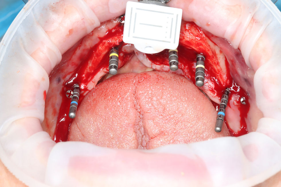
A central pin was placed to verify our position, and we proceeded with implant placement.
At sites 32 and 42, we placed 3.5 × 12 mm implants, both achieving excellent stability, with ISQ values of approximately 71–72.
We then placed tilted distal implants at sites 35 and 45, aiming to maximise the available implant length while respecting the emergence of the mental nerves.
Again, excellent stability was achieved.
There is, however, another small detail that illustrates the reality of a surgery of this magnitude.
The implant at site 45 ended up being 4 mm in diameter rather than the planned 3.5 mm.
When performing extensive procedures under general anaesthesia, we can plan everything down to the millimetre, but it is impossible to anticipate every intraoperative decision. We do not always have every possible combination of implants and prosthetic components immediately at hand.
When it came time to place the SRAs, we realised that we did not have the angled SRA we needed.
We had to borrow one from Clínica Maló.
It is a small detail, but it is also real-life Implant Dentistry.
On the radiograph, one implant can be seen tilted while the other is more upright. This created no difficulty in obtaining passivity, and we were able to structure the mandibular rehabilitation appropriately.
And now: to load or not to load?
At the end of surgery, we had an extremely stable mandibular situation.
The maxilla was a different story.
We had implants behaving very differently from one another and some sites where primary stability was less reassuring. The implant at 11 had even been submerged.
This was therefore a high-risk immediate loading situation.
We decided to proceed with a splinted rehabilitation, using the biomechanical advantage of rigidly connecting implants with different individual stability characteristics into a single functional unit.
Naturally, the patient also needs to understand that “immediate loading” does not mean “immediate unrestricted function.”
Our postoperative dietary instructions were therefore particularly strict.
The role of general anaesthesia
This procedure was performed under general anaesthesia.
We used local articaine in the maxilla primarily for its vasoconstrictive effect, both buccally and palatally.
This is particularly important in the palate. Good haemorrhage control during flap elevation significantly improves visibility of the surgical field.
General anaesthesia also provides some logistical advantages in procedures of this magnitude. The ability to administer intravenous medication, including corticosteroids and antibiotics when clinically indicated, can help optimise certain aspects of perioperative management.
Furthermore, while the patient was recovering from anaesthesia, we had several hours available.
And this ultimately influenced our prosthetic decision
.
Digital or conventional?
For the immediate loading procedure, we essentially had two possibilities.
The first was to continue with a fully digital workflow, using the cylinders associated with the surgical guide that we had developed through coDiagnostiX and Exocad.
The second was to do what we used to do in immediate loading cases several years ago: follow a conventional workflow.
A template in the mouth, intraoral connection of the components, impressions and laboratory work.
Because the patient would inevitably require several hours to recover from general anaesthesia, we decided to use that time.
We took the impressions and transferred the case to the laboratory.
The dental technician was able to carefully adapt the cylinders and complete the acrylic processing using a pressure pot.
Sometimes the most technologically sophisticated solution is not necessarily the solution that makes the most sense at that particular moment.
Being digital does not mean being dogmatic.
The real advantage of mastering a digital workflow is also knowing when we can return to a conventional technique without compromising predictability.
Immediate loading
Once the patient had recovered and we were back in the outpatient setting at IAD, we proceeded with delivery of the provisional restorations.
We verified adaptation and passivity and performed the necessary occlusal adjustments.
The patient had started the day with teeth so mobile that their movement was visible during a normal conversation.
She finished the day with a fixed maxillomandibular rehabilitation.
Her satisfaction was enormous.
We performed a postoperative maxillomandibular CBCT and scheduled the first postoperative appointment for seven days later.
Now another phase begins: allowing biology to do its work.
What this case taught me — again
This was not a perfect case.
And perhaps that is precisely why it is worth sharing.
The maxilla forced us to change positions, search for anchorage wherever bone was available, manage significant buccal deficiencies and, in one implant, simply accept that there was not enough stability to include it in the immediate loading protocol.
The mandible was considerably more predictable.
The prosthetic phase could have followed a more digital workflow, but the circumstances led us to choose a conventional approach.
And there was a third variable: execution.
Because perfect planning does not end at the computer.
The entire team needs to know exactly what has been planned, which implants are going to be used, what the alternatives are and which components will be required at each stage.
Ultimately, this case reminded me once again of three different dimensions of our profession:
planning, biology and execution.
Digital technology allows us to visualise, measure, anticipate and prepare.
The team allows us to execute what we have planned.
But once the flap is raised, biology still has the final say.
Perhaps that is the main message of this case:
We plan implants from the teeth. We execute the plan according to the available bone. And we change the plan whenever biology tells us to.

Comments