From Blurry to Brilliant: How CBCT is Rewriting Dental Diagnostics
- Andre Chen

- May 4, 2025
- 4 min read
The Magnificent World of Artificial Intelligence in Radiologic Images: The CBCT Revolution

Cone Beam Computed Tomography (CBCT) has emerged as a groundbreaking tool in the field of radiology, providing volumetric imaging with isotropic voxels and exceptional spatial resolution. This technology has revolutionized the way clinicians view and assess maxillofacial structures, offering detailed visualizations at significantly lower radiation doses compared to conventional CT scans.
At the core of its transformative power, CBCT integrates seamlessly into clinical workflows, enhancing diagnostic precision across various specialties. From implant planning and endodontics to orthognathic assessments, temporomandibular joint (TMJ) evaluations, and airway analysis, CBCT facilitates a deeper understanding of bone morphology, anatomical variations, and pathological conditions. This leads to evidence-based decision-making and improved surgical predictability, ensuring that clinicians can deliver more accurate and efficient care.
As a cornerstone of digital dentistry, CBCT empowers clinicians to provide safer, functionally driven care, enhancing both treatment outcomes and patient safety. However, there’s an even more exciting frontier to explore when it comes to CBCT—its integration with Artificial Intelligence (AI).
The AI-Driven CBCT: A New Era in Radiologic Imaging

I’ve had the privilege of watching this technology evolve over the years. From my time as an undergraduate at Lisbon University to my years at NYU, I’ve seen firsthand how CBCT technology has grown. I vividly remember the days when tomography was done with analog films, images only visible on a negatoscope or under direct sunlight. The shift to digital and 3D imaging has been nothing short of remarkable. In fact, I was fortunate to be part of the last class of Professor Humberto Ferreira da Costa and his team (including Dra Leonor de Sousa) in 1999, and watching the incredible progression since then has been amazing.
At the International Advanced Dentistry (IAD Lisbon), we made a conscious sacrifice to invest in state-of-the-art CBCT technology. This decision was made with one core objective in mind: to decrease radiation exposure while enhancing the precision of our diagnostic imaging. With this cutting-edge equipment, we are not only improving patient safety but also taking our diagnostic and treatment planning capabilities to new levels of accuracy and reliability
Artifact removal in CBCT is crucial for enhancing image clarity by reducing distortions caused by metal restorations, motion, or beam hardening. Advanced algorithms can now effectively minimize these artifacts, leading to more accurate diagnostics. Artificial intelligence (AI) further refines image quality by automatically detecting and correcting imperfections. AI also aids in segmentation, anatomical landmark identification, and pathology detection with greater speed and precision. Together, artifact reduction and AI elevate CBCT to deliver sharper, more reliable insights for clinical decision-making.

The Critical Role of High-Resolution Imaging

One of the most decisive factors in the clinical value of CBCT is its high-resolution imaging capability. The ability to capture fine anatomical details directly influences outcomes in several key areas:
Pathology Detection: High-resolution images allow for early and accurate identification of pathologies such as cysts, tumors, periapical lesions, and subtle bone defects. Detecting these conditions at their earliest stages can make a significant difference in prognosis and treatment success.

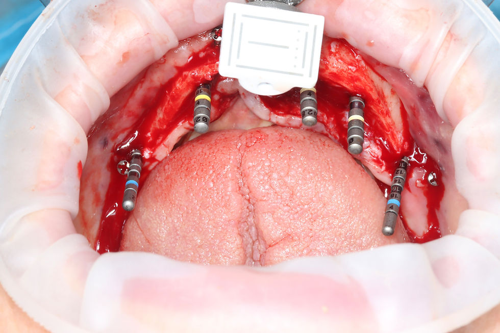
Implant Procedures: Precise visualization of bone density, morphology, and anatomical landmarks (such as the inferior alveolar nerve or maxillary sinus boundaries) is critical for safe and effective implant placement. High-resolution CBCT images reduce the margin of error, allowing for better planning and risk assessment.

Guided Surgery and Navigation Hardware: For computer-guided implant surgery and real-time navigation systems, the foundation is always high-quality, high-resolution CBCT data. Accurate imaging ensures that surgical guides fit perfectly and that navigation hardware can reliably track the planned surgical path. This directly translates into increased surgical precision, minimally invasive techniques, and improved patient outcomes.
When paired with AI, the value of high-resolution imaging is amplified even further. AI-enhanced algorithms can analyze these crystal-clear datasets to automatically detect anatomical structures and pathologies, providing an even higher level of diagnostic confidence.
From Diagnosis to Treatment: How AI and CBCT Are Changing the Game

The true power of the AI-CBCT partnership lies in its ability to assist clinicians in real-time decision-making. With AI continuously analyzing imaging data, it aids in the early detection of issues that might otherwise go unnoticed, leading to quicker interventions and more effective treatments. The integration of AI algorithms into CBCT not only speeds up the diagnostic process but also enhances the accuracy of interpretations, ensuring that clinicians have all the information they need to make informed decisions.
This technology doesn’t replace the expertise of radiologists or clinicians—it enhances it. AI in CBCT acts as an intelligent partner, providing insights that support more precise, personalized treatment planning. This collaboration redefines what’s possible in clinical imaging, pushing the boundaries of traditional diagnostic tools and offering a window into the future of intelligent healthcare.
The Future of CBCT and AI: A Glimpse into Tomorrow
As AI technology continues to evolve, the future of CBCT holds even greater promise. We are on the verge of refining imaging techniques, leveraging predictive analytics, and integrating data from multiple medical sources to offer truly holistic patient care. This will lead to a more seamless and connected healthcare experience, where diagnostic imaging is faster, more accurate, and more predictive.
We’re witnessing the dawn of a new era, where AI doesn’t replace radiologists but empowers them. CBCT, once a powerful tool on its own, is now an even more indispensable asset in the hands of clinicians, offering new dimensions of diagnostic and treatment possibilities.
Real-World Examples of CBCT in Action

As we continue to explore the potential of AI and CBCT, there are countless examples of how this technology is already making a difference. Whether it’s detecting subtle bone defects, identifying pathologies earlier, planning safer implant placements, or providing clearer images for guided surgical procedures, CBCT’s integration with AI is changing the way we practice medicine and dentistry.
In this magnificent world of artificial intelligence in radiologic images, CBCT stands as a testament to how technology can enhance the human touch in patient care. Together, AI and CBCT are not just improving clinical workflows—they’re shaping the future of healthcare.
Comments