
1
Immediate Implant Replacement of Adjacent Maxillary Central Incisors
Immediate implant placement in the esthetic zone with preservation of the buccal architecture
Immediate implants

Initial clinical presentation of a 40-year-old patient with severe internal and external root resorption affecting both maxillary central incisors.
THE CHALLENGE
A 40-year-old female patient, previously treated orthodontically and with an otherwise healthy dentition, presented with severe internal and external root resorption affecting both maxillary central incisors. CBCT assessment confirmed that neither tooth could be predictably preserved.
From a patient perspective, the first challenge was communicating the unexpected loss of two intact and highly visible anterior teeth in a young patient with no history of caries.
From a clinical perspective, however, the greater challenge was biological and esthetic. The simultaneous loss of both maxillary central incisors creates one of the most demanding scenarios for peri-implant soft-tissue management. Once two adjacent teeth are removed and replaced by implants, the periodontal ligament and its associated attachment apparatus are lost. Consequently, maintaining the original height and architecture of the interimplant papilla becomes significantly less predictable.
The primary concern from the outset was therefore not simply implant osseointegration or replacement of the missing teeth, but how to minimize changes to the facial and, particularly, interproximal tissue architecture when replacing two adjacent maxillary central incisors.
CLINICAL EXAMINATION
Clinical examination revealed preserved periodontal attachment around both maxillary central incisors, with no evidence of facial or interproximal attachment loss. Probing depths were approximately 2 mm facially and 3 mm interproximally.
Bleeding on probing was present and was more pronounced around tooth 21, particularly at the palatal and interproximal aspects, corresponding clinically to the area affected by the external resorptive process.
Despite the severity of the underlying root pathology, the facial and interproximal soft-tissue architecture remained largely preserved.
Importantly, the patient presented with a high smile line, making preservation of the existing gingival margins and papillary architecture particularly relevant to the esthetic outcome.
The patient was systemically healthy, a non-smoker, and reported no known drug allergies or relevant medication interactions. No systemic risk factors that could negatively influence healing or implant therapy were identified.
The combination of a high esthetic demand, preserved preoperative soft-tissue architecture, and the need to replace two adjacent maxillary central incisors made tissue preservation a central objective from the outset.


RADIOGRAPHIC EXAMINATION
CBCT examination of teeth 11 and 21 revealed extensive internal root resorption, with cavitation and palatal extension, associated with external root resorption affecting both maxillary central incisors.
Despite the severity of the root resorption, the surrounding alveolar anatomy remained remarkably preserved. The facial bone plate was intact at both sites, with no evidence of facial bone loss. Interproximal bone levels between the central and lateral incisors were also preserved.
No periapical pathology was identified at either site.
Following multidisciplinary assessment with the endodontic team, the extent and morphology of the resorptive defects were considered incompatible with predictable endodontic sealing and long-term preservation of the affected teeth. Extraction was therefore considered the most predictable treatment option.


TREATMENT PLANNING
The treatment strategy was based primarily on the integrity of the facial bone plate.
Within our clinical protocol, an intact facial plate in the anterior esthetic zone strongly favors consideration of immediate implant placement, provided that atraumatic extraction can be achieved, the residual socket anatomy is favorable, and adequate primary stability can be obtained.
The first critical step was therefore atraumatic extraction of teeth 11 and 21 while preserving the facial and interproximal socket walls. The definitive treatment pathway would only be confirmed following extraction and direct clinical assessment of both sockets.
Two scenarios were established preoperatively.
Plan A — Immediate implant placement
If the facial and interproximal walls remained intact following extraction and adequate primary stability could be achieved, two implants would be immediately placed using a fully guided static surgical protocol, followed by grafting of the facial peri-implant gaps and immediate fixed provisionalization.
Plan B — Delayed implant placement
If extraction resulted in significant compromise of the facial plate or the residual anatomy did not provide conditions for predictable immediate placement, the immediate protocol would be abandoned in favor of a staged approach.
A subtle preoperative facial volume discrepancy was identified at site 11 compared with site 21. For this reason, a palatal connective tissue graft at site 11 was incorporated into the treatment plan.
Immediate provisionalization was planned from the outset. Two provisional crowns were digitally designed and CAD-CAM manufactured, with the intention of intraorally picking them up onto temporary cylinders following implant placement.
The provisional restorations were therefore conceived not merely as temporary teeth, but as an integral component of the peri-implant tissue-management strategy from the first day of healing.


Evidence Box 1
Is immediate implant placement predictable in an intact anterior extraction socket?
Clinical Translation
TheWISE Concept®
01 THE QUESTION
Can two adjacent maxillary central incisors be replaced with immediate implants while minimizing the inevitable changes to the interproximal soft-tissue architecture?
02 THE DECISION
Following atraumatic extraction of teeth 11 and 21, direct inspection confirmed preservation of the facial and interproximal socket walls at both sites.
The decision was therefore made to proceed with two immediately placed implants, facial peri-implant grafting, and non-occlusal immediate loading with individual implant-supported provisional restorations.
Rather than considering immediate placement solely as a surgical opportunity, the protocol was selected as a means of minimizing the biological, esthetic and social impact of losing two maxillary central incisors in a young patient with a high smile line.
The objective was to transition, whenever biologically possible, from:
Natural tooth → Implant → Fixed provisional restoration
within a single surgical intervention, while maintaining continuous support of the existing soft-tissue architecture.
03 THE WISE RATIONALE
W — Whole
The treatment was planned around the patient as a whole rather than around two extraction sockets.
She was young, socially active and presented with a high smile line. Losing both maxillary central incisors represented not only a biological and esthetic challenge, but also a potentially significant social burden.
Whenever the anatomy allowed it, minimizing the period during which the patient experienced the consequences of tooth loss was therefore an important treatment objective.
I — Individual
The protocol was individualized to the patient's specific anatomy and circumstances.
Following atraumatic extraction, the facial and interproximal socket walls remained intact, allowing the immediate strategy to proceed.
A single-stage approach also reduced the need for multiple surgical interventions, prolonged treatment phases, and removable or adhesive provisional solutions.
S — Science
The decision was supported by the biological principles and available evidence for immediate implant placement in appropriately selected anterior extraction sockets, provided that adequate primary stability, correct three-dimensional implant positioning and appropriate management of the peri-implant gap can be achieved.
The biological limitation associated with replacing two adjacent teeth was nevertheless acknowledged: loss of the periodontal attachment apparatus inevitably alters the interproximal tissue architecture.
The objective was therefore not to prevent remodeling, but to control its magnitude.
E — Execution
The biological and patient-centered rationale was translated into a precise clinical protocol:
Atraumatic extraction → Fully guided immediate implant placement → Xenogeneic grafting of the facial peri-implant gaps → Site-specific soft-tissue augmentation → Non-occlusal immediate loading → Controlled emergence profile → Definitive ceramic rehabilitation.
SURGICAL PROCEDURE
Local anesthesia was administered using articaine with epinephrine 1:100,000 facially and 1:200,000 palatally.
Both maxillary central incisors were removed using a strictly atraumatic, flapless extraction protocol. No full-thickness mucoperiosteal flap was elevated, with the objective of preserving the vascular supply to the facial and interproximal periodontal tissues.
Following extraction, direct inspection confirmed preservation of the socket walls and allowed the immediate protocol to proceed as planned.
Implant positioning had been planned through a completely digital workflow. A preoperative intraoral scan was merged with the CBCT dataset in coDiagnostiX, and a tooth-supported surgical guide was manufactured for a fully guided Straumann surgical protocol.
Two Straumann TLX NT 3.35 × 10 mm implants with a 1.8-mm machined collar were placed through the surgical guide in prosthetically driven positions.
The machined collar was deliberately selected to provide a polished transmucosal interface around which the grafted hard and soft tissues could mature, while minimizing the risk of future exposure of a rough implant surface.
Primary stability was assessed using resonance frequency analysis, with ISQ values of approximately 71–72 at sites 11 and 21, supporting the decision to proceed with immediate loading.
The facial jumping gaps were grafted with a xenogeneic bone substitute, carefully packed while maintaining the existing socket architecture.
Because site 11 presented a subtle preoperative facial volume deficiency relative to site 21, a small connective tissue graft was harvested from the palate and positioned facially at site 11. The graft was stabilized using non-resorbable polyamide sutures.
Postoperative pharmacological management included antibiotic therapy, a non-steroidal anti-inflammatory drug (NSAID), and paracetamol-based analgesia as required. Systemic corticosteroids were not routinely prescribed for this type of surgical procedure.


Evidence Box 2
What happens to the interimplant papilla when two adjacent anterior teeth are replaced by two implants?
Clinical Translation
DIGITAL LAB WORK
The digital workflow began with a preoperative intraoral scan acquired using CEREC, combined with CBCT data obtained with a Planmeca Viso 7.
The intraoral and CBCT datasets were merged in coDiagnostiX, allowing prosthetically driven three-dimensional planning of both implants.
Two Straumann TLX NT 3.35 × 10 mm implants with a 1.8-mm machined collar were selected.
Implant depth and three-dimensional position were planned in relation to the existing alveolar anatomy and the intended restorative outcome. Particular attention was given to the relationship between implant position, the machined collar, the restorative components and the intended emergence profile.
The patient's preoperative tooth morphology was used as the reference for provisional restoration design. Two provisional maxillary central incisors were designed in exocad, with the objective of reproducing as closely as possible the original tooth position, proportions and morphology.
Rather than attempting to finalize the implant–provisional relationship entirely in the virtual environment, the digitally designed provisional restorations were subsequently intraorally picked up onto temporary cylinders after implant placement.
This allowed the final emergence profiles to be adapted to the actual three-dimensional implant positions and the clinical soft-tissue conditions encountered at surgery.
The provisional restorations were subsequently finished and polished, with particular attention to their transmucosal contours.
Digital planning defined the restorative destination; intraoral adaptation allowed the emergence profile to respond to the actual biology.


PROSTHETIC PROCEDURE
Two temporary cylinders were connected directly to the implants, and the preoperatively designed provisional crowns were intraorally picked up onto the cylinders.
The emergence profiles were adjusted, finished and polished to provide appropriate peri-implant tissue support while simultaneously helping to contain the xenogeneic graft within the facial peri-implant gaps during early healing.
Both provisional restorations were immediately loaded, but were carefully adjusted to remain completely free of centric and eccentric occlusal contacts.
This non-occlusal immediate loading protocol allowed the patient to receive fixed implant-supported restorations on the day of surgery while minimizing functional loading during the early osseointegration phase.
The screw-access channels were sealed with PTFE tape and composite resin.
At approximately 10–14 days, the surgical sites demonstrated uneventful healing, with healthy soft tissues and preservation of the intended peri-implant architecture.
Following four months of healing and soft-tissue maturation, the case progressed to the definitive restorative phase.
At this stage, the objective was not to redesign the emergence profile, but rather to accurately transfer the tissue architecture created and maintained during provisionalization into the definitive restorations.
A digital impression was obtained using an intraoral scanner with two implant-level scan bodies directly connected to the implants.
Two individual screw-retained zirconia crowns veneered with feldspathic ceramic were fabricated.
Particular attention was given to reproducing the contours established by the provisional restorations, thereby maintaining the peri-implant tissue support developed throughout the healing period.
The laboratory achieved excellent integration of shade, morphology, surface texture and characterization with the adjacent natural dentition.
The definitive restoration should inherit the tissue architecture created by the provisional—not force the tissues to adapt to a new contour.


FINAL OUTCOME
At the six-month follow-up, the outcome was favorable from both patient-centered and clinical perspectives.
From a Whole-patient perspective, the patient was highly satisfied with the final result. The loss of both maxillary central incisors was managed within a single surgical intervention, without a prolonged period involving removable appliances or adhesive provisional restorations.
Clinically, comparison between the initial and six-month situations demonstrated the biological remodeling that had been anticipated from the outset.
The most evident change occurred interproximally between implants 11 and 21, where a reduction in papillary height was observed. Consequently, the definitive central incisors required slightly longer and more rectangular cervical proportions than the original natural teeth.
This tissue change did not compromise the patient's perception of the esthetic result but illustrates an important biological limitation of replacing two adjacent natural teeth with two implants:
The immediate protocol minimized interproximal tissue collapse, but could not completely prevent it.
At six months, further soft-tissue maturation should still be expected; the current result should therefore not be interpreted as the definitive long-term tissue position.
An unexpected finding at site 11
An additional observation concerned the facial soft-tissue augmentation performed at site 11.
Despite placement of a connective tissue graft, the final facial volume at site 11 was not greater than that observed at the contralateral site 21. Clinically, site 11 demonstrated more remodeling than initially anticipated, suggesting substantial remodeling or loss of the volume originally provided by the connective tissue graft during healing.
This did not compromise the rehabilitation. Nevertheless, it raises a clinically relevant question regarding the routine indication for connective tissue grafting in otherwise favorable immediate implant sites.
If an additional surgical procedure increases patient morbidity but does not necessarily translate into a clinically meaningful long-term volume benefit, its indication should be considered site-specific rather than automatic.
Biological stability
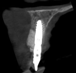
Radiographically, both implants demonstrated favorable osseointegration and stable peri-implant conditions.
Of particular importance was the maintenance of the facial tissue complex around the implant platform in the region of the grafted peri-implant gap.
In a young patient, the objective cannot be limited to obtaining an attractive six-month photograph. These implants may be expected to function for several decades, making long-term biological stability, maintainability and preservation of peri-implant tissue volume at least as important as the immediate esthetic result.
This case therefore demonstrates both the potential and the limitations of immediate implant therapy for two adjacent maxillary central incisors.
Success should not be judged by whether remodeling occurred, but by whether its magnitude was controlled while achieving a stable, maintainable and patient-acceptable result.




CLINICAL PEARLS
1. Plan for different outcomes — and choose the protocol you can execute predictably.
Treatment planning should anticipate the different clinical scenarios that may occur before and during surgery.
When more than one technique can provide a comparable biological and esthetic outcome, the preferred strategy should be the one that offers the greatest predictability in the clinician's own hands.
Evidence defines what is possible; experience and reproducibility help determine what is appropriate for the individual case.
2. More surgery does not necessarily mean better treatment.
Every additional surgical intervention increases morbidity, treatment time and the potential for complications.
Additional procedures—including soft-tissue augmentation—should therefore have a clear biological or clinical indication rather than being incorporated routinely into a protocol.
If an additional surgery does not provide a meaningful expected benefit, its necessity should be questioned.
3. Treat the whole patient, not the missing tooth.
The clinical significance of losing a tooth extends beyond the extraction socket.
Smile line, function, social interaction, expectations, tolerance for removable or adhesive provisional restorations, treatment duration and willingness to undergo multiple surgical procedures can all influence the optimal treatment strategy.
The same anatomical defect may therefore require different solutions in different patients.
The most sophisticated treatment is not necessarily the one with the most procedures—it is the one that achieves the required outcome with the least biological and personal cost.
CASE CREDITS
Surgical Treatment: Prof. André Chen
Prosthetic Rehabilitation: Dr. João Borges
Digital Workflow: Dr. Sofia Hernández
Dental Laboratory: António Louro
LEARN MORE
Readers interested in exploring the biological principles behind this case should consider four areas of evidence:
Immediate implant placement
Survival, complications, case-selection criteria and esthetic outcomes following immediate implant placement in the anterior maxilla.
Adjacent implants and interproximal tissue architecture
Interimplant bone dimensions, papillary height and the biological differences between tooth–implant and implant–implant interfaces.
Immediate implant-site biology
Healing of the facial peri-implant gap, dimensional changes of the facial bone and the potential role of grafting procedures.
Provisionalization and emergence profile management
The role of immediate provisional restorations in supporting peri-implant tissues during healing and transferring the developed tissue architecture to the definitive restoration.
Understanding the biological limitations of immediate implant therapy is as important as understanding its surgical possibilities.